| SOMATIC
CELL SAMPLING AND RECOVERY (SCSR™) |
 SCSR™
is a technology for the
preservation, transport, and recovery of cells from fecal samples. In
the late 1980's, our founder Dr. Padmanabhan P. Nair, then at a major US government
laboratory (USDA-ARS),
sought a biopsy-free method to obtain target cells and genetic material for the large-scale human nutrition studies
that he was conducting. Over the next
decade the cell isolation system and the preservative solution went through a
series of improvements, starting from a complex countercurrent centrifugal
elutriation (1989), to a freeze-thaw
linear density gradient
fractionation (1991), and finally resulting in the current
room
temperature step-gradient system (1999), which Dr. Nair developed after retirement.
Numerous formulations were evaluated for use in the transport medium, which enables
samples to be collected off-site and transported to the lab with minimal cell
degradation. Dr. Nair continued his research, receiving support from the Small
Business Innovation Research Program (SBIR) which resulted in two human
studies on colorectal cancer and inflammatory bowel disease. The SCSR system is now available to the medical and scientific
community in a simple kit format comprising sampling tubes with transport media,
filters and strainers, and step-gradient cushion used for the separation of the cells in a
standard swinging-bucket non-refrigerated centrifuge.
SCSR™
is a technology for the
preservation, transport, and recovery of cells from fecal samples. In
the late 1980's, our founder Dr. Padmanabhan P. Nair, then at a major US government
laboratory (USDA-ARS),
sought a biopsy-free method to obtain target cells and genetic material for the large-scale human nutrition studies
that he was conducting. Over the next
decade the cell isolation system and the preservative solution went through a
series of improvements, starting from a complex countercurrent centrifugal
elutriation (1989), to a freeze-thaw
linear density gradient
fractionation (1991), and finally resulting in the current
room
temperature step-gradient system (1999), which Dr. Nair developed after retirement.
Numerous formulations were evaluated for use in the transport medium, which enables
samples to be collected off-site and transported to the lab with minimal cell
degradation. Dr. Nair continued his research, receiving support from the Small
Business Innovation Research Program (SBIR) which resulted in two human
studies on colorectal cancer and inflammatory bowel disease. The SCSR system is now available to the medical and scientific
community in a simple kit format comprising sampling tubes with transport media,
filters and strainers, and step-gradient cushion used for the separation of the cells in a
standard swinging-bucket non-refrigerated centrifuge.
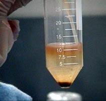
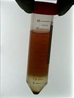
The technique involves collecting
0.5 gm (a small pellet) of stool in the tube provided (containing a sampling
scoop and a preservative) and
transporting the specimen to the laboratory under normal ambient temperature within 5
days. The suspension is filtered, underlayered with a dense cushion medium,
and centrifuged at low speed for 10 minutes. Cells are extracted from the interface between the
supernatant and cushion (the pellet, which contains clumped, dense, or
debris-bound cells, is retained as a backup). The cells are washed repeatedly
in PBS, aliquoted, and are ready for analysis or archiving. They can be stained for specific cell surface markers using immunofluorescent antibodies
and subjected to flow cytometry. They can be lysed for the extraction of proteins,
glycoproteins, mRNA, and genomic DNA.
- Transport
specimen without refrigeration up to 5 days after collection
- Typically yields several million viable cells from a small sample size (0.5-1.0 g)
- Minimizes problem of bacterial interference,
Taq inhibition, and fluorescence quenching
- All cell types
are isolated
- Nontoxic components in the test kit
- Procedure requires only a table-top centrifuge (50-ml tube) and common laboratory supplies
- Cells can be cultured, cells can be archived for years
The
gastrointestinal tract is the most toxic environment in the body. The
primary way the body deals with this fact is by cellular attrition: the entire epithelium of the gastrointestinal tract is renewed every 5-7 days, with different rates at different sites (eg stomach vs. small intestine vs. colon). The source of the replacement cells are stem cells
residing at or near the base of the gastrointestinal crypts. In the case of the colon the cells are generally thought to be at the base. There are multiple feedback mechanisms, still unexplained, that regulate the rate of stem cell division and the differentiation pathways. This is an area of current research.
As the stem cells divide, at least one daughter cell remains undifferentiated. As cell division progresses toward the top of the crypts (the colon does not have villi), the cells differentiate into intermediate transit cells (also called progenitors) that eventually become functional terminally differentiated cells. The
major functional cells of the colon are the absorptive columnar cells and the mucous-producing goblet cells. After reaching the epithelial surface the cells are shed into the fecal stream or phagocytosed. (Although from our work on culturing exfoliated cells, it appears that some shed cells retain or revert to their undifferentiated or partially differentiated character).
The epithelium of the colon contains billions
of cells. Hence many millions of cells are replaced daily. From our work it is clear that
large numbers of these cells survive fecal passage and retain viability.
 Somatic cells isolated by this
technology represent an important source of informational macromolecules providing a picture of the immediate past history of various organ systems and their
functioning. Inflammatory processes, autoimmune disorders, host response to
pharmaceuticals, mutational events and such other conditions can be investigated
by examining these cells. Some examples of potential
applications are: BIOCHEMISTRY, CANCER RESEARCH, RHEUMATOLOGY,
IMMUNOLOGY (MUCOSAL IMMUNITY), MEDICINE, GASTROENTEROLOGY, MOLECULAR AND CELLULAR BIOLOGY,
NUTRITION, SOMATIC CELL GENETICS, PHARMACOGENOMICS.
Somatic cells isolated by this
technology represent an important source of informational macromolecules providing a picture of the immediate past history of various organ systems and their
functioning. Inflammatory processes, autoimmune disorders, host response to
pharmaceuticals, mutational events and such other conditions can be investigated
by examining these cells. Some examples of potential
applications are: BIOCHEMISTRY, CANCER RESEARCH, RHEUMATOLOGY,
IMMUNOLOGY (MUCOSAL IMMUNITY), MEDICINE, GASTROENTEROLOGY, MOLECULAR AND CELLULAR BIOLOGY,
NUTRITION, SOMATIC CELL GENETICS, PHARMACOGENOMICS.
Copyright©2000-2008 NonInvasive Technologies, LLC. All rights
reserved